BD Biosciences T-Cell Research
BD Biosciences 대리점, 서린바이오사이언스 입니다.

T-Cell Subtypes cytotoxic T cells / helper T cells(Th) / regulatory T cells(Tregs)
| Type of Cell | Cytotoxic | Th1 | Th2 | Th95 | Th17 | Tfh6 | Treg |
| Main Function | Kill virus-infected cells | Activate microbicidal
function of |
Help B cells and switch antibody isotype production | T cell proliferation and neutrophil response production by B cells | Enhance neutrophil
response |
Regulate development of antigen specienhanced IgG and IgE Enhance fic B cell development and antibody production | Immune regulation |
| Extracellular Markers | CD8 | CD4 CXCR3 | CD4 CCR4, Crth2 (human) | CD4 | CD4, CCR6 | CD4, CXCR5 | CD4, CD25 |
| Differentiation
Cytokines |
IFN-g, IL-2, IL-12, IL-18, IL-27 | IL-4, IL-2, IL-33 | IL-4,TGF-b | TGF-b, IL-6, IL-1, IL-21, IL-23 | IL-12, IL-6 | TGFb- , IL-12 | |
| Effector Cytokines | IFN-g,TNF, LT-a | IFN-g, LT-a,TNF | IL-4, IL-5, IL-6, IL-13 | IL-9, IL-10 | IL-17A, IL-17F, IL-21, IL-22, IL-26 ,TNF, CCL20 | IL-21 | TGF-b, IL-10 |
| Transcription Factors | T-bet,Stat1,Stat6 | GATA3,Stat5,Stat6 | GATA3,Smads,Stat6 | RORgt, RORa, Stat3 | Bcl-6, MAF | FoxP3,Smad3,Stat5 |
– To support the use of multicolor flow cytometry for the study of T cells, BD offers a deep portfolio of reagents, which are highlighted in pink.
T-Cell Research Platforms
| Cell Surface Markers
: To Identify cells from heterogeneous samples |
[Phenotyping of Cells with Unique Surface Profiles]
T cells and their subsets can be identified by differential expression of cell surface markers including CD3, CD4, CD8, CD25, CD127, and CD196 (CCR6).
Adding markers such as CD197 (CCR7), CD62L, CD69, and CD45RO to an analysis provides important information about the potential for cells to home and
localize within the body, as well as the activation status of the T-cell subset of interest. This information can also be used to identify different memory subsets.
With the availability of multiple BD Horizon Brilliant Violet and Brilliant Ultraviolet dyes, larger panels can be created that include multiple dim markers.
Rich data sets can be obtained from precious samples. The 13-color panel below examines memory and activation status of multiple T-cell subsets.
| Marker | Fluorochrome | Purpose |
| Viability dye | V500 | Viability |
| CD3 | PerCP-Cy5.5 | T cell marker |
| CD4 | BUV395 | T cell subsetting |
| CD8 | FITC | T cell subsetting |
| CD127 | Alexa Fluor® 647 | RegulatoryTCell Marker |
| HLA-DR | APC-H7 | Activation |
| CD45RO | PE-Cy7 | Memory |
| CD197 (CCR7) | BV421 | Naïve/Memory |
| CD38 | BV605 | Activation |
| CD27 | BV786 | Memory |
| CD25 | PE-CF594 | Regulatory T Cell
Marker/Activation |
| CD196 (CCR6) | PE | Th17 Cell Marker |
| CXCR3 | Alexa Fluor® 700 | Th1 Cell Marker |
| Intracellular flow cytometry
: A powerful tool for the study of T-cell differentiation |
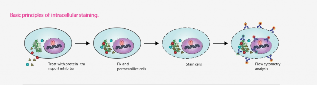
| BD Cytofix/Cytoperm™
: fixation/permeabilization solution – 다양한 Cytokines 및 Cell surface marker staining 목적으로 사용 |
BD Pharmingen™
: transcription factor buffer set – Transcription factor alone or Cell surface marker, cytokines과 혼합되어 있는 Transcription factor staining 목적으로 사용 |
BD Phosflow™
: permeabilization buffer III – Phosphoepitope 검출을 위한 Flow cytometry 분석 시 Permeabilization 목적으로 사용 |
| BD™ Cytometric Bead Array (CBA)
: To measure secreted cytokines within a sample |
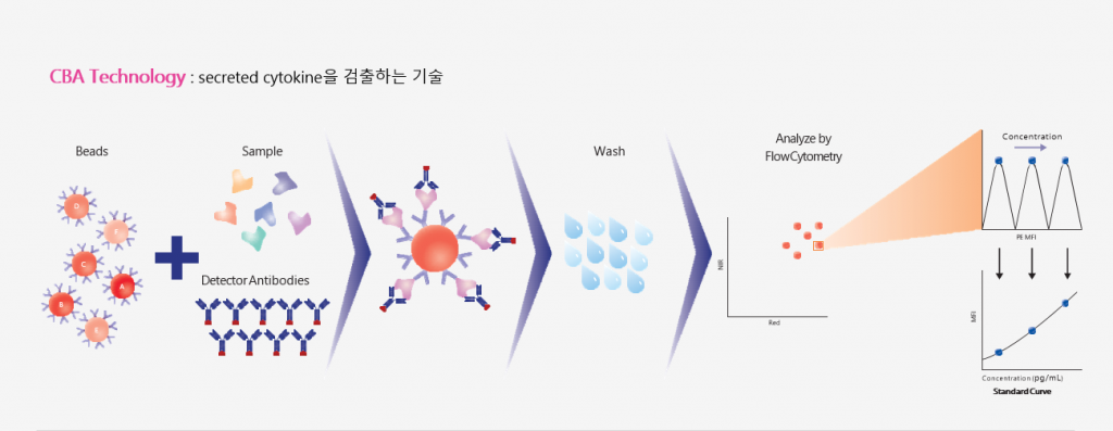
CBA products 장점
– 여러 cytokine을 동시에 분석 가능 (multiplexing)
– 소량의 샘플로도 Quantitative Result 획득 가능
– 타사 제품 대비 분석 소요 시간이 짧음
*CBA vs. ELISA vs. ICS 비교 *intracellular cytokine staining (ICS)
| CAPABILITY | CBA | ELISA | ICS |
| Allows detection of multiple cytokines in same experiment | ✓ | ✓ | |
| Can obtain phenotype of specific cells expressing cytokine of interest | ✓ | ||
| Can measure quantity of cytokine secreted | ✓ | ✓ |